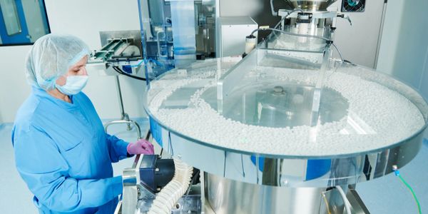
Discovery
Our discovery services are designed to accelerate the path from concept to breakthrough. Leveraging state-of-the-art technologies and a team of seasoned scientists, we offer a comprehensive suite of services including molecular biology, genomics, protein science, and immunology. Our approach integrates cutting-edge methodologies with deep scientific expertise to uncover novel insights and drive innovation. From initial hypothesis generation and experimental design to data analysis and interpretation, we provide end-to-end support to ensure robust and reproducible results. Our collaborative model fosters close partnerships with our clients, enabling customized solutions tailored to their unique research needs and goals. Whether you are in academia, biotechnology, or the pharmaceutical industry, our research discovery services are committed to pushing the boundaries of bioscience and transforming your innovative ideas into tangible discoveries.

Research & Development
Our assay method development services are designed to create precise, reliable, and reproducible assays tailored to your specific research and development needs. Leveraging advanced technology and a team of experienced scientists, we offer comprehensive solutions that encompass everything from initial assay conceptualization and design to optimization and validation. We specialize in developing a wide array of assays, including biochemical, cell-based, immunoassays, and high-throughput screening assays, ensuring robust and sensitive detection of target analytes. Our meticulous approach includes extensive testing and validation to meet regulatory standards and achieve high-quality results. We work closely with our clients to understand their unique requirements and challenges, enabling us to deliver customized assay solutions that accelerate drug discovery, development, and commercialization. Whether for academic research, biotechnology, or pharmaceutical applications, our assay method development services are committed to driving scientific advancement and innovation.

Manufacturing
We leverage our extensive network and industry expertise to identify and engage reliable contract manufacturers in the U.S., Asia, and Europe. This global reach allows us to offer customized solutions tailored to meet diverse production needs and quality standards. Our dedicated team works closely with clients to manage the entire process, from initial scouting and vetting of manufacturers to final production and quality assurance. By providing end-to-end support, we enable our clients to scale efficiently and effectively, ensuring that their products meet market demands while maintaining the highest standards of quality and compliance.
Cell and Gene Therapy Custom Services
Custom GMP Protein
As the potential of cellular therapies continues to expand, the demand for high-quality raw materials and ancillary components, such as GMP cytokines and growth factors, increases. Our extensive supply of GMP proteins is supported by our commitment to providing cell therapy manufacturers with a consistent, safe, and traceable supply of reagents. Develop a new protein or produce your existing version under GMP and Animal-Free quality standards required for use as ex vivo cell therapy reagents.

Custom Antibody
We offer comprehensive custom antibody services designed to meet your specific research needs. Our team of experts utilizes advanced technologies and rigorous quality control measures to develop high-affinity, high-specificity antibodies tailored to your targets. From antigen design and peptide synthesis to hybridoma development and monoclonal antibody production, we provide end-to-end solutions that ensure reliable and reproducible results. Whether you require antibodies for diagnostic, therapeutic, or research applications, our custom services deliver exceptional performance and support to advance your scientific goals. Partner with us to experience unparalleled expertise and innovation in antibody development.

Stem Cell culture media
Our Custom Cell Culture Media Manufacturing and Services team collaborates with you to expedite and standardize media production, develop optimized formulations, and conduct specialized media testing. Our capabilities encompass media and supplement production, GMP media manufacturing, media formula optimization, custom labeling, and assays for testing media on stem cells, immune cells, and other cell lines. We leverage our expertise to ensure high-quality, reproducible results tailored to your specific research and production needs.

Custom immunoassay development
we tailor assays to meet your specific research and diagnostic needs. Our team of experts utilizes cutting-edge technologies and extensive industry experience to design, develop, and validate highly sensitive and specific immunoassays. Whether you need enzyme-linked immunosorbent assays (ELISA), multiplex assays, or other immunoassay formats, we provide end-to-end solutions, including antigen design, antibody production, assay optimization, and validation. Our comprehensive approach ensures robust, reproducible results that advance your scientific and clinical objectives. Partner with us to benefit from bespoke immunoassay solutions that deliver exceptional performance and accuracy.

IVD Antibody Development Services
Monoclonal Antibody Development
Recombinant Antibody Development
Monoclonal Antibody Development
Monoclonal antibodies are produced from a single clone of B cells and are highly specific to a single epitope on an antigen. They are known for their high specificity and consistency, making them ideal for diagnostic assays.
Process:
- Antigen Preparation: Selection and preparation of the target antigen.
- Immunization: Immunizing mice or other host animals with the prepared antigen.
- Cell Fusion: Fusing spleen cells from the immunized animal with myeloma cells to create hybridoma cells.
- Screening: Screening the hybridoma cells for those producing the desired antibody.
- Cloning: Cloning positive hybridoma cells to ensure monoclonality.
- Production: Culturing the cloned cells to produce monoclonal antibodies.
- Purification: Purifying the antibodies from the culture supernatant.
- Characterization: Characterizing the antibodies for specificity, affinity, and other relevant properties.
Polyclonal Antibody Development
Recombinant Antibody Development
Monoclonal Antibody Development
Polyclonal antibodies are produced by different B cell clones in response to an antigen, recognizing multiple epitopes. They offer higher sensitivity and broader detection capabilities for various applications.
Process:
- Antigen Preparation: Selection and preparation of the target antigen.
- Immunization: Immunizing host animals (e.g., rabbits, goats) with the prepared antigen.
- Booster Immunizations: Administering booster shots to enhance antibody production.
- Serum Collection: Collecting blood from the immunized animals.
- Antibody Extraction: Extracting antibodies from the serum.
- Purification: Purifying the antibodies to remove non-specific proteins.
- Characterization: Characterizing the antibodies for specificity, affinity, and other relevant properties.
Recombinant Antibody Development
Recombinant Antibody Development
Recombinant Antibody Development
Recombinant antibodies are generated through genetic engineering, allowing for precise control over the antibody sequence and structure. These antibodies can be designed for improved specificity and stability.
Process:
- Gene Synthesis: Synthesizing the genes encoding the antibody variable regions.
- Cloning: Inserting the antibody genes into expression vectors.
- Expression: Transfecting suitable host cells (e.g., CHO cells) with the expression vectors.
- Screening: Screening for high-expressing clones.
- Production: Culturing the selected clones to produce recombinant antibodies.
- Purification: Purifying the antibodies from the culture supernatant.
- Characterization: Characterizing the antibodies for specificity, affinity, and other relevant properties.
Antibody Pairs Development
Antibody Fragment Development
Recombinant Antibody Development
Antibody pairs are essential for sandwich assays such as ELISA and immunochromatographic tests, where one antibody captures the antigen and the other detects it. These pairs must be carefully selected and validated for optimal performance.
Process:
- Antigen Preparation: Selection and preparation of the target antigen.
- Antibody Generation: Developing monoclonal or polyclonal antibodies against the antigen.
- Pair Screening: Screening antibody pairs for their performance in sandwich assays.
- Optimization: Optimizing the selected pairs for maximum sensitivity and specificity.
- Validation: Validating the antibody pairs in relevant diagnostic assays.
- Production: Producing and purifying the validated antibody pairs.
- Characterization: Characterizing the antibody pairs for performance parameters such as sensitivity, specificity, and stability.
Antibody Humanization
Antibody Fragment Development
Antibody Fragment Development
Antibody humanization involves modifying non-human antibodies (e.g., mouse antibodies) to resemble human antibodies more closely. This process reduces the immunogenicity of the antibodies when used in human therapeutic applications.
Process:
- Sequence Analysis: Analyzing the sequence of the original non-human antibody.
- Human Framework Selection: Selecting suitable human antibody frameworks.
- Grafting CDRs: Grafting the complementarity-determining regions (CDRs) from the non-human antibody onto the human frameworks.
- Back-Mutation: Introducing critical back-mutations to restore the original binding affinity.
- Expression: Cloning the humanized antibody genes into expression vectors and transfecting them into host cells.
- Screening: Screening for clones that express the humanized antibody.
- Production: Culturing the selected clones to produce the humanized antibodies.
- Purification: Purifying the antibodies from the culture supernatant.
- Characterization: Characterizing the antibodies for specificity, affinity, immunogenicity, and other relevant properties.
Antibody Fragment Development
Antibody Fragment Development
Antibody Fragment Development
Antibody fragments, such as single-chain variable fragments (scFv) and single-domain antibodies (VHH), offer several advantages, including smaller size and better tissue penetration. They are useful in both diagnostic and therapeutic applications.
Process:
- Antigen Preparation: Selection and preparation of the target antigen.
- Phage Display Library Construction: Constructing a phage display library expressing antibody fragments.
- Library Screening: Screening the phage display library to identify antibody fragments that bind to the target antigen.
- Clone Selection: Selecting positive clones expressing high-affinity antibody fragments.
- Expression: Cloning the selected antibody fragment genes into expression vectors and transfecting them into host cells.
- Production: Culturing the host cells to produce antibody fragments.
- Purification: Purifying the antibody fragments from the culture supernatant.
- Characterization: Characterizing the antibody fragments for specificity, affinity, stability, and other relevant properties.
IVD Kit Development
ELISA Kit Development
Lateral-Flow Immunochromatographic Assay (LFIA) Kit Development
RadioImmunoAssay (RIA) Kit Development
Enzyme-Linked Immunosorbent Assay (ELISA) kits are widely used for detecting and quantifying antigens or antibodies in various samples. They are known for their high sensitivity, specificity, and ease of use in both research and clinical diagnostics.
Process:
- Antigen/Antibody Selection: Identifying and selecting the appropriate antigen or antibody for the assay.
- Coating: Coating microplate wells with the antigen or antibody.
- Blocking: Blocking non-specific binding sites to reduce background noise.
- Sample Addition: Adding samples to the wells, allowing the target analyte to bind to the coated antigen/antibody.
- Detection Antibody: Adding a detection antibody conjugated with an enzyme to the wells.
- Substrate Addition: Adding a substrate that the enzyme converts to a detectable signal, typically a color change.
- Signal Measurement: Measuring the signal using a microplate reader.
- Validation: Validating the kit for sensitivity, specificity, precision, and reproducibility.
- Packaging: Packaging the kit with all necessary reagents and instructions for use.
RadioImmunoAssay (RIA) Kit Development
Lateral-Flow Immunochromatographic Assay (LFIA) Kit Development
RadioImmunoAssay (RIA) Kit Development
Radioimmunoassay (RIA) kits use radioactively labeled substances to measure the concentration of antigens or hormones in samples. RIAs are highly sensitive and can detect very low levels of analytes.
Process:
- Antigen/Antibody Selection: Identifying and selecting the appropriate antigen or antibody for the assay.
- Radiolabeling: Labeling the antigen or antibody with a radioactive isotope.
- Sample Preparation: Preparing samples for analysis.
- Incubation: Incubating samples with radiolabeled antigen/antibody and an unlabeled antigen/antibody to allow competitive binding.
- Separation: Separating bound from free antigen/antibody using precipitation or other methods.
- Measurement: Measuring the radioactivity of the bound fraction using a gamma counter.
- Standard Curve: Creating a standard curve to determine the concentration of the analyte in the samples.
- Validation: Validating the kit for sensitivity, specificity, precision, and reproducibility.
- Packaging: Packaging the kit with all necessary reagents and instructions for use.
Lateral-Flow Immunochromatographic Assay (LFIA) Kit Development
Lateral-Flow Immunochromatographic Assay (LFIA) Kit Development
Latex Particle-Enhanced Turbidimetric Immunoassay (LETIA) Kit Development
Lateral-flow immunochromatographic assay (LFIA) kits, also known as rapid tests, are used for point-of-care testing. These kits provide quick and easy-to-read results, making them suitable for various diagnostic applications.
Process:
- Antigen/Antibody Selection: Identifying and selecting the appropriate antigen or antibody for the assay.
- Conjugate Preparation: Conjugating antibodies with colored particles (e.g., gold nanoparticles).
- Strip Assembly: Assembling the test strip with sample pad, conjugate pad, nitrocellulose membrane, and absorbent pad.
- Sample Application: Applying the sample to the test strip.
- Capillary Action: Allowing the sample to flow through the strip by capillary action, where it interacts with the conjugate and the test/control lines.
- Result Interpretation: Interpreting the test result based on the appearance of lines on the strip.
- Validation: Validating the kit for sensitivity, specificity, precision, and reproducibility.
- Packaging: Packaging the kit with all necessary components and instructions for use.
Latex Particle-Enhanced Turbidimetric Immunoassay (LETIA) Kit Development
Latex Particle-Enhanced Turbidimetric Immunoassay (LETIA) Kit Development
Latex Particle-Enhanced Turbidimetric Immunoassay (LETIA) Kit Development
Latex particle-enhanced turbidimetric immunoassay (LETIA) kits use latex particles to enhance the detection of antigens or antibodies in samples. The formation of antigen-antibody complexes causes an increase in turbidity, which is measured to determine the analyte concentration.
Process:
- Antigen/Antibody Selection: Identifying and selecting the appropriate antigen or antibody for the assay.
- Latex Conjugation: Conjugating the antigen or antibody to latex particles.
- Reagent Preparation: Preparing reagents and buffers for the assay.
- Sample Addition: Adding samples to a reaction cuvette containing the latex reagent.
- Reaction: Allowing the antigen-antibody reaction to occur, forming complexes that increase turbidity.
- Measurement: Measuring the turbidity using a turbidimeter or spectrophotometer.
- Standard Curve: Creating a standard curve to determine the concentration of the analyte in the samples.
- Validation: Validating the kit for sensitivity, specificity, precision, and reproducibility.
- Packaging: Packaging the kit with all necessary reagents and instructions for use.
Immunohistochemistry (IHC) Kit Development
Latex Particle-Enhanced Turbidimetric Immunoassay (LETIA) Kit Development
Immunohistochemistry (IHC) Kit Development
Immunohistochemistry (IHC) kits are used for the detection of specific antigens in tissue sections. They are widely used in pathology to diagnose diseases and to study the distribution and localization of biomarkers.
Process:
- Antigen Retrieval: Preparing tissue sections and retrieving antigens using heat or enzyme treatment.
- Blocking: Blocking non-specific binding sites to reduce background staining.
- Primary Antibody: Applying the primary antibody specific to the target antigen.
- Secondary Antibody: Applying a secondary antibody conjugated with an enzyme or fluorescent dye.
- Detection: Using chromogenic substrates for enzyme-conjugated antibodies or direct visualization for fluorescent antibodies.
- Counterstaining: Counterstaining the tissue sections to highlight cell structures.
- Microscopy: Examining the stained tissue sections under a microscope.
- Validation: Validating the kit for sensitivity, specificity, precision, and reproducibility.
- Packaging: Packaging the kit with all necessary reagents and instructions for use.
Copyright © 2019-2024 Nelson Labs - All Rights Reserved.
This website uses cookies.
We use cookies to analyze website traffic and optimize your website experience. By accepting our use of cookies, your data will be aggregated with all other user data.